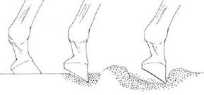
: A hard surface with high impact resistance does not allow the toe to dig in during push off. Centre: A surface with moderate impact and shear resistance allows the toe to dig but then offers resistance as the hoof pushes off. Right: A soft surface with low impact but low shear resistance and gives way and does not offer sufficient resistance as the hoof pushes off.
JOINTS
Joints will deteriorate with age and also the workload of the horse, especially if the surfaces they are worked on are too hard, the workload is too demanding when the horse is young and still developing.
Support for Ageing Joints
With age the cartilage starts to “dry out”, it can be compared to a kitchen sponge that becomes brittle when dry, but becomes soft and spongy again when it is wet. Well cartilage needs water to stay spongy too, however the process of keeping that water (hydrolysing) needs glucosamine. That is converted to GAGs and they cause the hydrolysing of the cartilage. With age the horses natural levels of glucosamine drop and so the sponge dries out and the cushioning effect of the joint reduces and the horse loses its spring. It then proceeds onto degenerative joint condition – osteoarthritis.
Joint Damage, Injuries, Trauma
The other main source of joint degeneration comes from trauma – the concussive damage from the repetitive pounding of the feet and legs on firm surfaces. There is help at hand, these conditions can be alleviated by correctly formulated nutriceuticals that contain the right amounts of scientifically proven actives (such as collagen , glucosamine, chondroitin and manganese), injectable therapies from your veterinarian, helpful shoeing and care to check surfaces for working the horse and good nutrition with balanced healthy diet containing correct amounts of minerals.
The Equine Hock Joint
Dr Peter Gillespie. BVSc MACVS.
Situated midway between the stifle joint and the foot in the hind limb, is the hock, one of the hardest working joints in the equine body. It is also one of the most complex – comprising six bones making up four individual joints, all held in place by numerous ligaments and joint capsules
The largest joint of the four is the tibiotarsal joint – the articulation between the tibia and the talus. The three smaller hock joints in descending order are the proximal inter-tarsal, distal inter-tarsal and tarso-metatarsal joints.
For all practical purposes, the hock works as a hinge, moving by flexion and extension through one plane. Practically all of the movement occurs in the tibiotarsal joint. Movement in the other joints is minimal, restricted by the shape of the articular surfaces of the bones themselves, the collateral ligaments and the strong fibrous joint capsule. A special anatomical arrangement exists between the stifle and the hock, which allows them to work in synchrony with each other – when the stifle flexes, the hock flexes, when one extends, so does the other.
All equine disciplines require full and free flexion of both the stifle and hock joints to achieve effective hind limb propulsion. Whether it is the acceleration necessary in racing or the collection of dressage, the hock is the pivotal hind limb joint. Even at slow gaits, huge stresses are placed on the hock. Although the hind legs are not subjected to the same concussive forces of weight bearing as the front legs, the loading on them during movement is still significant. Shock waves travel up the limb and through the bones, ligaments and joint capsules that collectively make up the hock. In addition, the break over phase of the stride produces a rotational force (torque) that is also absorbed by the structures of the hock. The absorption of these forces is the reason why the hock is the most common site in the hind limb of work (stress) related injuries.
Good conformation is important in minimising the stress forces. Conversely, poor conformation exacerbates the stress even during low intensity work.
In assessing conformation, it is important to view a horse standing square, on an even, flat surface. From the rear view, normal hock conformation should feature a straight axis through the tibia (gaskin) and cannon bone, with no deviation at the hock. Bearing in mind that most horses ‘toe out’ slightly behind, it is easy to get the wrong impression of them being cow hocked. From the side view, normal hocks feature a vertical cannon bone with an angle to the tibia of close to 150°.
Cow hocks are a common conformational fault. When viewed from the rear, there is a deviation at the hock from the axis of the gaskin to the cannon bone. This conformation puts additional stress on the medial structures and predisposes those horses to bone spavin (refer later).
Sickle hocks are less common but when they do occur they usually place strain on the plantar ligament at the back of the hock, resulting in a condition commonly known as a curbed hock (refer later).When viewed from the side, sickle hocks feature the cannon bone angled forward from the vertical axis.
Straight hocks occurs when a horse has very little angulation between the thigh bone (femur) and the tibia (gaskin). Good hock angulation is a desirable conformational trait in all horses. Straight hocks prevent a horse from reaching as far forward with the hind legs during propulsion.
Bowed hocks are the opposite of cow hocks. They are not a common conformational fault but when they do occur they put excessive strain on the outside structures of the joints.
Hock lameness occurs when the stresses placed on the hock joints produce inflammatory changes which interrupt normal structure and function. The initial signs of lameness can be so subtle that they are often not seen as being related to a hock problem. Usually the first noticeable sign is stiffness associated with muscle soreness in the lumber region of the back. Poorly trained chiropractors (of which there are many) earn a good income from telling owners their horse’s ‘back is out’ when in fact they have a simple secondary muscle soreness from a primary hock problem. Because the lumbo-sacral joint in the lower back flexes in unison with the stifle and hock joints, any restriction in the mobility of the hock will affect the lower back as well.
Another consistent early sign of hock lameness is pain around the head of the medial (inside) splint bone. Because it contributes a large weight bearing part of the tarso-metatarsal joint, excessive strain on the joint will produce referred pain in the upper part of the ligament of the splint bone. The test to detect this pain is known as the ‘Churchill Hock Test’ named after veterinarian who first showed the pain to be a consistent early feature of hock lameness.
More often than not these early signs go undetected or mis-diagnosed. Swellings of the joint capsule or a boney enlargement on the inside of the hock are often the first noticeable signs. By this stage there is inflammation in the joint capsule, degeneration of the joint cartilage, and remodeling of the underlying bone.
There are several specific hock conditions that are worth mentioning.
Bone Spavin
This is another colloquial term used to describe a distension of the joint capsule of the tibiotarsal joint. It presents as a soft fluctuating swelling on the inside front aspect of the hock, with another smaller swelling slightly higher on the outside. If finger pressure is applied to either one of these swellings, the other can be felt to enlarge.
Although the name implies a specific hock condition, bog spavin should be treated as a symptom of an underlying hock problem. More often than not, such problems are nothing more than a slight strain of the joint capsule with mild inflammation but no associated lameness. Occasionally there is a more serious joint problem and lameness, that requires veterinary attention. Osteochondritis dissecans (OCD) is a common cause of bog spavin in young horses.
Bog Spavin
This is another colloquial term used to describe a distension of the joint capsule of the tibiotarsal joint. It presents as a soft fluctuating swelling on the inside front aspect of the hock, with another smaller swelling slightly higher on the outside. If finger pressure is applied to either one of these swellings, the other can be felt to enlarge.
Although the name implies a specific hock condition, bog spavin should be treated as a symptom of an underlying hock problem. More often than not, such problems are nothing more than a slight strain of the joint capsule with mild inflammation but no associated lameness. Occasionally there is a more serious joint problem and lameness, that requires veterinary attention. Osteochondritis dissecans (OCD) is a common cause of bog spavin in young horses.
Thoroughpin
This is a distension of the tendon sheath that encloses the deep digital flexor tendon at the back of the hock. While not essentially a condition of the hock joint, it has a similar appearance to a bog spavin and therefore, needs to be distinguished from it. The swelling associated with thoroughpin is higher up and further back than a bog spavin and may be pressed from inside to outside and vice versa. Thoroughpin does not usually cause lameness and is generally regarded as of little significance.
Curbed Hock
The plantar tarsal ligament is a broad ligament that runs down the back of the hock. Sprain of this ligament is not uncommon particularly in horses with sickle hock conformation. Although the ligament appears swollen and is usually painful to the horse when palpated, lameness is not usually a feature.
Capped Hock
Kicking out at either stall walls or float doors can cause a haematoma on the point of the hock. Blood accumulates beneath the unbroken skin and can take several weeks to dissipate. Drainage should be avoided because of the complications that can result.
Septic Arthritis
An infection in the tibiotarsal joint can have a similar appearance to a bog spavin. The difference is, the horse is usually acutely lame. Often there is an obvious sign of a puncture wound however this may not always be the case, particularly in foals. This is a potentially crippling condition that requires immediate veterinary attention.
The Equine Suspensory Ligament
Dr Peter Gillespie. BVSc MACVS.
Injuries to the suspensory ligament are a common occurrence in athletic horses. They can occur in both the fore and hind legs and have the potential to bring a horse’s competitive career to an end.
Where is the suspensory ligament and what does it do? To describe it in simple terms, it runs down behind the cannon bone between the knee and the fetlock in the fore leg and between the hock and the fetlock in the hind leg.
To be more precise, in the fore leg it originates on the distal row of carpal bones at the back of the knee and on the back of the upper part of the metacarpus (cannon bone). In the hind leg, it originates mainly on the upper metatarsus, although there are some attachments to the distal row of tarsal (hock) bones.
Two thirds of the way down the metacarpus (or metatarsus) it divides into medial and lateral branches which continue down to attach to the outside surface of the sesamoid bones at the fetlock joint. From there it continues below the fetlock as lateral and medial extensor branches which insert on the Common Digital Extensor tendon at the front of the pastern between the fetlock and the foot.
It is interesting that the suspensory ligament is actually a modified muscle. It’s anatomical equivalent in animals with more than one toe is the medial interosseous muscle. In the horse the suspensory ligament is made up of predominately tendon fibres with some residual muscle fibres. The number of muscle fibres varies between individual horses and between breeds. Standardbred horses have a higher proportion than thoroughbreds.
The suspensory ligament along with the sesamoid bones and distal sesamoidean ligaments make up what is known as the suspensory apparatus. Its function is to support the fetlock joint during the weight-bearing phase of the stride.
It is during this phase that most suspensory ligament injuries occur. Uneven loading of the limb during weight bearing is the main contributing cause helped in many cases by an uneven ground surface and poor foot balance. Overloading of the ligament leads to tearing of collagen (tendinous) fibres and the small blood vessels associated with the muscle fibres. There is bleeding within the ligament with the formation of a haematoma.
The healing process proceeds through three steps;
(1) Removal of damaged tissue by phagocytes (white blood cells).
(2) Migration of fibroblasts into the area to start producing new collagen (scar tissue).
(3) Remodelling of scar tissue.
The early scar tissue is organized in a haphazard manner. During the first 2-3 months the collagen fibres orientate themselves in a parallel alignment and slowly increase in diameter.
The repaired tissue is not as strong or as elastic as a normal ligament tissue and as such is predisposed to re injury. This is an important point when assessing the prognosis for a successful return to competition.
The inflammatory changes associated with the tearing of the collagen fibres produce the characteristic signs of heat, swelling, pain and reduced function. The term for inflammation of the suspensory ligament is desmitis. We recognize six distinct conditions.
1. Avulsion of the origin of the suspensory ligament
This condition usually affects the forelimbs. It involves a tearing of the attachment of the ligament at the back of the metacarpus. Signs can vary from an acute, severe lameness to a chronic recurring lameness that can be difficult to pinpoint. Swelling is not always obvious because the ligament at this point is surrounded on three sides by bone (cannon & two splint bones). A common feature of lameness associated with this type of injury is that it is usually worse when the horse is trotted in a circle with the injured leg to the outside. Diagnosis is usually dependent on nerve blocks, ultrasonography and radiography Treatment involves box rest for the first 4-6 weeks followed by 6-8 months paddock rest. The prognosis is good for a return to competition without recurrence of the injury.
2. Proximal suspensory desmitis
affects the suspensory in the uppermost quarter. It is a common cause of both fore and hindleg lameness. Again there is often no swelling with this condition due to the surrounding boney structures. Signs can vary greatly from a pronounced lameness to un-diagnosed poor performance. Lameness can be exacerbated by either trotting in a circle with the affected leg to the outside or by flexion of the fetlock joint. Diagnosis can be confirmed by nerve blocks and ultrasonography. The preferred treatment is a combination of box rest and controlled exercise. Box rest is necessary for the first 4-6 weeks to complete the initial phase of the healing process. Six months of controlled exercise helps the remodeling process by stimulating better alignment of the collagen fibres. The prognosis with proximal suspensory injuries varies with the age of the injury prior to diagnosis. Acute injuries are more likely to respond to treatment with a successful return to competition than are chronic injuries.
3. Desmitis of the suspensory body
The body of the suspensory is defined as the section between the upper proximal quarter and the bifurcation to the medial and lateral branches. This injury is more common in the fore legs. Swelling is generally a feature, as is pain on palpation. Lameness is usually not an early sign, in fact nine times out of ten swelling will precede any lameness. Because the body of the suspensory ligament lies close to the narrower diameter sections of the splint bones, swelling in the ligament can put pressure on these bones, causing them to fracture. For this reason it is advisable to take radiographs when this type of injury occurs. Again initial box rest and anti-inflammatory therapy followed by controlled exercise is the preferred treatment. If a splint bone is fractured it should be removed surgically. The prognosis with this type of injury is guarded.
4. Desmitis of the branch of the suspensory ligament
This is the easiest suspensory injury to diagnose because of the obvious swelling that fills the natural hollow between the ligament and the cannon bone. The swollen branch is always painful on palpation. Lameness is usually not a feature. Box rest, anti-inflammatory therapy and controlled exercise are important in the recuperative phase but the prognosis with this type of injury is poor.
5. Suspensory desmitis secondary to splints or fractured splint bones
Splints are a common occurrence in young horses. Besides being a source of pain, they can encroach on the adjacent ligament causing a focal desmitis. Often it is the less obvious splints (blind splints) that cause problems.
6. Suspensory Breakdown Injuries
Complete failure of the suspensory apparatus occurs from time to time as a high speed injury in thoroughbred racehorses and eventers. It is due to acute over-loading of the support structures of the fetlock during the weight bearing phase at the gallop. Either the suspensory ligament or the sesamoid bones break down depending on the fitness of the horse. The fitter the horse, the more likely it will break its sesamoid bones. This suggests that the suspensory ligament strengthens with training. There is an acute, severe lameness with this condition with dropping of the fetlock joint. Salvage for breeding is the only option.
Apart from avulsion injuries, the chance of a full recovery with a return to competition is generally poor. During the first 10-14 days post injury it is important the horse be confined and aggressive anti-inflammatory therapy instigated. This should consist of the early application of cold packs and bandaging combined with systemic medication. By keeping the initial inflammatory reaction to a minimum, the amount of damaged tissue that has to be later removed and remodeled is reduced.
Joints – Damage, Arthritis, DJD
What is Equine Arthritis & Degenerative Joint Disease
Every Step They Take
Arthritis means inflammation in the horse’s joints, this inflammation can be from any cause: infection, trauma etc. However Degenerative Joint Disease is a disorder, common in animals and humans alike usually referred to as DJD, it is often used as an overall definition for all joint disorders, although there are various forms of arthritis and different reasons for the development of such.
It is generally regarded as a non-inflammatory condition of articular cartilage and is often called osteoarthritis. There are two classifications: primary DJD- which occurs where there is no known direct trauma or disease but is typical of the joint changes in the older animal; secondary DJD- from direct trauma or infection of the joint. Secondary DJD and its inevitable progression to inefficient joints can occur at any age and several factors may start the disorder. Sadly once DJD starts, it cannot be reversed or cured. However the process can be slowed & relief can be provided that may result in a horse maintaining an active life. DJD is the number one reason for premature horse retirement, 60% of all lameness is due to DJD. Joints are lined by specialised tissue, called hyaline cartilage that is vital to smooth joint function. This cartilage can become frayed and damaged due to the mechanical wear, ultimately exposing bone and causing pain. With more chronic cases, the soft tissue supporting structures can become thickened which results in a decreased range of motion of the joint. DJD is not a condition of articular cartilage alone, the synovial membrane which lines the joint cavity, is also affected. If left unattended joints will become swollen and sore and eventually new bone is created to strengthen the surface (a process called sclerosis) and extend its margins (bone spurs and osteophytes). In its final stages, if left unchecked, arthritis causes the fusing of equine joints. The key is the extent of the progress and acting early so that it can be held at a point that the horse is pain free and still useful symptoms can be alleviated.
What Causes The Damage to the Joints?
The main cause is trauma, either a direct one off or repetitive concussive forces, the most common is the latter, that is the force up the leg from each step the horse takes. The likelihood of DJD comes from: poor confirmation, genetic pre-disposition, surfaces too hard, overworking on harder surfaces, poor shoeing, direct accidental damage to a joint, infection of a joint, puncture of a joint or old age.
How Can DJD be Avoided
So to prevent the onset of DJD, start from the earliest days and provide good balanced nutrition to maximise quality bone and joint development, before breaking in a young horse should be assessed for conformation defects. If a career is planned for racing, jumping, dressage etc., then an experienced veterinarian should assess whether the conformation is strong and correct enough for the chosen career. Over taxing an unsuitably built horse is likely to produce DJD and therefore a decision not to pursue a particular path may mean that given an easier sport, DJD and pain for the horse can be delayed or prevented. Good intelligent shoeing can assist, but a farrier should not try and correct a deviation of the leg as that will only cause stress on joints which otherwise may not be susceptible. Good support with balanced level feet and correct angles are vital to assist prevention, on the other hand, incorrect shoeing can often be a trigger. Ongoing he needs to be well shod, to reduce stress and concussive forces to the joints, shoes should be selected for the conditions of work, ie heavy road shoes should not be used unless really necessary and the horse is strong enough to carry the weight. Care with the development of the young horse, not breaking in too early, balancing the workload with the development of maturity while being aware of the type of horse and its future. Larger heavier warmbloods mature much later than thoroughbreds. Excessive workload can be a cause, but the key factor is the surface the horse is worked on, trauma from concussion is the most common cause of DJD in younger horses. The idea of trotting horses out on roads to “harden them up”, is typical of the lack of understanding of what brings a horse to an early end of its competitive life. If DJD develops, early recognition can make a big difference to preventing progression.
The Importance of the Surface Under the Horse’s Feet
The desire to have a workable all weather surface, whether it be an arena or track, that is level and firm throughout the year, has resulted in hard sand or cinders etc. being used with no thought to cushioning the strike of the leg. Many are built as though they were a road and a shallow sand surface is placed on top. To avoid impact resistance, some thought must be given to cushioning that surface if the horse is not to start the negative processes towards DJD. Overseas surfaces are inherently deeper, softer, and sand being added to rubber, soft chips, plastic, or now fibre sand is utilised. The hoof moves forwards downwards, and rapidly decelerates when brought in contact with the ground. It’s this deceleration and strike and causes the concussive effect which can be damaging to joints and bones. The term impact resistance describes the ability of the footing to absorb that concussive effect, therefore hard surfaces have high impact resistance.
Sheer resistance describes the ease with which the footing is displaced by a shearing (rotational) force. When the leg is pushing against the ground to generate propulsion, the toe tends to rotate into the surface. The shear resistance of the footing should be low enough to allow the toe to dig in as the hoof pushes against it, reducing tension in the distal check ligament and reducing pressure of the deep digital flexor tendon on the navicular region. The shear resistance can be too low, eg deep soft dry sand (as above the tideline at the beach), the ground does not offer sufficient resistance to the hoof pushing against it. Instead, the surface gives way during push off and the muscles have to work harder to generate propulsion. As a consequence of having to work harder, the muscles can become fatigued more quickly and this predisposes the horse’s ligaments and tendons to injuries. Surfaces that are deep and soft will have low impact resistances but very deep heavy soft sand may be detrimental to ligaments and tendons as they will fatigue quicker with the effort of moving the legs through such a surface.
The negative effect of deep sand can be reduced by adding water which will improve the shear resistance as the foot moves through the sand, for example being ridden along the edge of the waterline on a beach is an improvement from the deeper dry sand and it still can provide a softer surface than hard dry sand. The answer lies in the middle, with low impact resistance, as suggested from using wood, fibre, rubber and other synthetics etc. hard and/or dry sand is the worst option and most likely to initiate the process of DJD. The depth of the hoof print is a good indicator of the impact resistance, the deeper the hoof print, the lower the impact resistance and the concussive effect transmitted to joints and bones.
Scientific studies actually show that impact force is much greater with sand, and can be dampened (reduced in force), by the addition of water or wood or fibre. Three levels of force were classified, the greatest being dense hard (asphalt), surfaces with friction damping (sand), the least being surfaces with structural damping (wood fibres). Whilst the cheapest option maybe hard shallow sand, it may be the most expensive if it shortens the active life of the joints of the horse! When veterinarians check a horse for lameness, they run it on a hard surface as the horse is much more likely to show lameness on such a surface ….. Go figure! Direct trauma to the joint can often develop into DJD, but any injury to a joint, strains, sprains, direct impact should be investigated and monitored so that the possibility of DJD is noted before it becomes chronic.
What are the Signs of Joint Pain?
At first the horse may not show actual lameness, initially the usual pain signs of discomfort, ears back, grumpy reaction to movement, restless tail when moving, personality change, then shortened steps, horse feels wooden. It then moves on into stronger reactions to pain, refusing to go, negative responses even when handled. Sadly some of the early signs are often missed with inexperienced owners/trainers suggesting the horse be forced, often with the whip. Early on there may be a mild intermittent lameness. It gets better with a turn out, but returns and increases with work. If the joint has a capsule that is distensible then some swelling and maybe heat is seen and felt. But if the joint is one that is contained by strong ligaments and or tendons, then swelling may not be evident. Early attention to the signs is so important as DJD can be ameliorated and held at a minor level.
To obtain a diagnosis a veterinarian may carry out a nerve block. After the area is located, X-rays will be taken. Also taking a sample of the fluid from the joint (synovial) can be tested to indicate the presence of arthritis. This can indicate the extent of the degeneration by the count of the cartilage and bone cells. The use of x-rays alone can create misleading diagnosis, in the early part of the disease very little sign of DJD may show on x-ray, some changes may show and could mis-lead the diagnosis, so x-rays are used after other signs are confirmed, to assess the extent of the disease, or other issues which may be the true cause of the pain. Arthroscopy is an option, usually done by a specialist, where an optical tube is inserted into the joint and gives the veterinarian a view of the joint and its issues. Changes can be seen as an area of dullness on the cartilage, colour change from glistening white to a mottled grey or even yellow, it gives the most definitive diagnosis of arthritis. However this is an invasive procedure and has all the risks of that invasion.
What Can be Done to Help a Horse with DJD
It is not curable, it is progressive, but it can be held, of course in the first instance the best we can do is to prevent it, as discussed above. A quick response to the signs is important.
Scientific studies have now shown that using a nutraceutical containing the correct active ingredients in correct quantities does have a recognised beneficial effect on joints showing signs of DJD, and that they help to maintain the quality of the cartilage and retain its elasticity and shock absorbing qualities. Ideally a nutriceutical should be used prior to DJD developing, especially with horses that may be vulnerable for any of the reasons given above. Prophylactic use can assist where a horse is required to work at a young age, as with racing. The nutriceutical must have certain active ingredients, at the right levels proven by scientific tests. For example the recommended level of glucosamine is 1200 mg per day, but manganese is needed in the formula to convert that to an active called GAGS as that is the substance that improves the cartilage. Perna Mussel, while effective in other animals and humans is not so effective for horses and a high quantity is needed. The newest science on a supplement is the exciting addition of Denatured Collagen Type II which in conjunction with the other proven actives of glucosamine and chondroitin has increased the therapeutic response.
For established DJD, injections directly into the joint, in combination with a suitable nutriceutical, like Flex-Equine Plus, has also been scientifically proven to reduce the negative reactions to DJD. Indeed this combination has been proven to reduce the number of injections required, and therefore the cost. Likewise intra- muscular injections of products like pentosan in combination with the same high standard nutriceutical can have similar benefits for horses not quite at such a level of the disease. Varied lengths of paddock rest can prolong the active use of the horse. Overall it becomes a maintenance regime to reduce the level of discomfort and achieve some extension of the horse’s competitive years. The main solution is to prevent the onset and safeguard joints with the next generation Flex-Equine Plus – protect the younger horse from concussive damage and put back the spring in the older horse! It has the right amounts of scientifically proven ingredients its a full formulation of glucosamine, chondroitin, manganese, gelatin, zinc and copper and now with newly discovered Undenatured Collagen Type II.
Reference’s for sources for this article:
M Cruz DVM, MVM, Msc, DrMedVetA.J Lipowitz, C.D. Newton E.Barrey, B. Landjerit, R Wolter Ecole Nationale Veterinaire d’Alfori France, Laboratoire de Biomecanique de L’ENSAM R.Lamberski, R A Lobos, Dr. D J Burba Dr. H M Clayton BVMS, PhD, Diplomate Michigan State University.
Some of the scientific references (for a fuller list see the product page Flex Equine Plus)
Therapeutic efficacy of undenatured type-II collagen (UC-II) in comparison to glucosamine and chondroitin in arthritic horses.Gupta RC, Canerdy TD, Skaggs P, Stocker A, Zyrkowski G, Burke R, Wegford K, Goad JT, Rohde K, Barnett D, DeWees W, Bagchi M, Bagchi D. Jvet Phamacol -2009
The Effect of Glucosamine and Chondroitin on Stressed Equine Cartilage Explants. R.S.A Harlan MS, R.C.A Haut PhD, M.W.A. Orth PhD JNL Equine Science Volume 32 Ussue 1 Jan 1012
Effects of Chondroitin and Glucosamine Sulfate in a Dietary Bar Formulation on Inflammation,0 Interleukin-1CEB2, Matrix Metalloprotease-9, and Cartilage Damage in Arthritis. M. Chou*,Nathalie Vergnolle ,Jason 0 J. McDougall,John L. Wallace,Stephanie Marty,Val Teskey and AndreG. Buret. Exp Biol Med April 2005 Vol 230 No 4
Effects of glucosamine hydrochloride and chondroitin sulphate, alone and in combination, on normal and interleukin-1 conditioned equine articular cartilage explant metabolism. Dechant JE, Baxter GM, Frisbie DD, Trotter GW, McIlwraith CW.
Equine Vet Jnl 2005 May 37 (3) :227-31


